Embark on a captivating journey into the realm of microscopy with our labeled diagram of a compound light microscope. This essential tool empowers scientists and researchers to unravel the intricate details of the microscopic world, unlocking a wealth of knowledge and understanding.
As we delve into the inner workings of this remarkable instrument, we’ll dissect its components, unravel the principles of illumination and magnification, and uncover the techniques for preparing and examining specimens. Join us as we unveil the secrets of the microscopic universe.
Microscope Overview: Labeled Diagram Of A Compound Light Microscope

A compound light microscope is a powerful tool that allows scientists to observe and study small objects. It consists of two or more lenses, the objective lens and the eyepiece, which combine to produce a magnified image of the specimen.
Compound light microscopes are used in a wide variety of applications, including biology, chemistry, and medicine. They are used to study cells, bacteria, viruses, and other small organisms. They can also be used to examine materials such as minerals, metals, and plastics.
The basic components of a compound light microscope include the following:
- Eyepiece: The eyepiece is the lens that the user looks through. It magnifies the image produced by the objective lens.
- Objective lens: The objective lens is the lens that is closest to the specimen. It gathers light from the specimen and focuses it on the eyepiece.
- Stage: The stage is the platform on which the specimen is placed. It can be moved up and down to focus the specimen.
- Condenser: The condenser is a lens that focuses light on the specimen. It helps to improve the quality of the image.
- Diaphragm: The diaphragm is a disk with a hole in the center. It controls the amount of light that reaches the specimen.
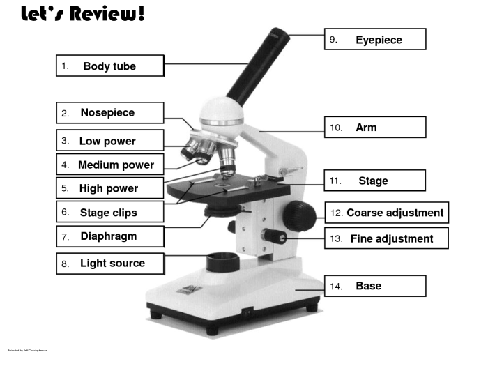
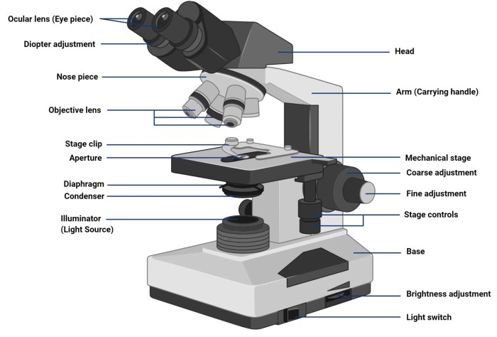
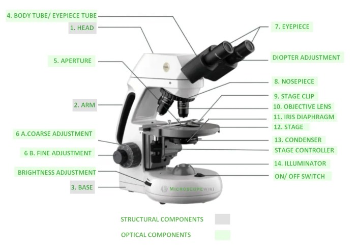
Microscope Diagram, Labeled diagram of a compound light microscope
The following diagram shows the labeled components of a compound light microscope:

- 1. Eyepiece
- 2. Objective lens
- 3. Stage
- 4. Condenser
- 5. Diaphragm
- 6. Light source
Illumination System
The illumination system of a compound light microscope consists of a light source, a condenser, and a diaphragm. The light source provides the light that illuminates the specimen. The condenser focuses the light on the specimen. The diaphragm controls the amount of light that reaches the specimen.
There are two main types of light sources used in compound light microscopes: incandescent light and LED light. Incandescent light sources are less expensive than LED light sources, but they produce more heat. LED light sources are more expensive than incandescent light sources, but they produce less heat and have a longer lifespan.
The condenser is a lens that focuses light on the specimen. It helps to improve the quality of the image by reducing glare and scattering. The diaphragm is a disk with a hole in the center. It controls the amount of light that reaches the specimen.
The size of the hole in the diaphragm can be adjusted to control the brightness of the image.
Objective Lenses
Objective lenses are the lenses that are closest to the specimen. They gather light from the specimen and focus it on the eyepiece. Objective lenses are available in a variety of magnifications, ranging from 4x to 100x. The magnification of an objective lens is determined by its focal length.
The shorter the focal length, the higher the magnification.
The numerical aperture (NA) of an objective lens is a measure of its ability to gather light. The higher the NA, the more light the objective lens can gather. The NA of an objective lens is determined by its refractive index and its angle of acceptance.
The resolving power of an objective lens is its ability to distinguish between two closely spaced objects. The resolving power of an objective lens is determined by its NA and the wavelength of light. The shorter the wavelength of light, the higher the resolving power.
The following table summarizes the magnification and NA of different objective lenses:
| Magnification | NA |
|---|---|
| 4x | 0.10 |
| 10x | 0.25 |
| 20x | 0.50 |
| 40x | 0.65 |
| 100x | 1.25 |
Eyepiece
The eyepiece is the lens that the user looks through. It magnifies the image produced by the objective lens. Eyepieces are available in a variety of magnifications, ranging from 10x to 20x. The magnification of an eyepiece is determined by its focal length.
The shorter the focal length, the higher the magnification.
The field of view of an eyepiece is the area that can be seen through the eyepiece. The field of view is determined by the magnification of the eyepiece and the size of the objective lens. The higher the magnification of the eyepiece, the smaller the field of view.
The depth of field of an eyepiece is the range of distances within which the specimen can be focused. The depth of field is determined by the magnification of the eyepiece and the NA of the objective lens. The higher the magnification of the eyepiece, the smaller the depth of field.
Focusing System
The focusing system of a compound light microscope allows the user to focus the specimen. There are two main types of focusing systems: coarse focusing and fine focusing.
Coarse focusing is used to bring the specimen into approximate focus. It is done by moving the stage up and down. Fine focusing is used to fine-tune the focus. It is done by moving the objective lens up and down.
The focusing system of a compound light microscope is essential for obtaining a clear and sharp image of the specimen.
Stage and Specimen Preparation
The stage of a compound light microscope is the platform on which the specimen is placed. It can be moved up and down to focus the specimen. There are two main types of stages: mechanical stages and motorized stages.
Mechanical stages are moved by hand. They are less expensive than motorized stages, but they are also less precise.
Motorized stages are moved by a motor. They are more expensive than mechanical stages, but they are also more precise.
Specimen preparation is an important step in microscopy. It involves preparing the specimen so that it can be viewed under the microscope. Specimen preparation techniques vary depending on the type of specimen being viewed.
Some common specimen preparation techniques include:
- Fixing: Fixing is a process that preserves the specimen. It can be done using chemicals or heat.
- Staining: Staining is a process that adds color to the specimen. It can help to make the specimen easier to see.
- Sectioning: Sectioning is a process that cuts the specimen into thin slices. It can help to make the specimen easier to view.
FAQ Insights
What is the purpose of a condenser in a compound light microscope?
The condenser focuses light onto the specimen, providing optimal illumination and enhancing the contrast of the image.
How does the numerical aperture of an objective lens affect its resolving power?
The higher the numerical aperture, the greater the resolving power, allowing for the visualization of finer details in the specimen.
What is the difference between coarse and fine focusing?
Coarse focusing brings the specimen into approximate focus, while fine focusing makes precise adjustments to achieve a sharp and clear image.

